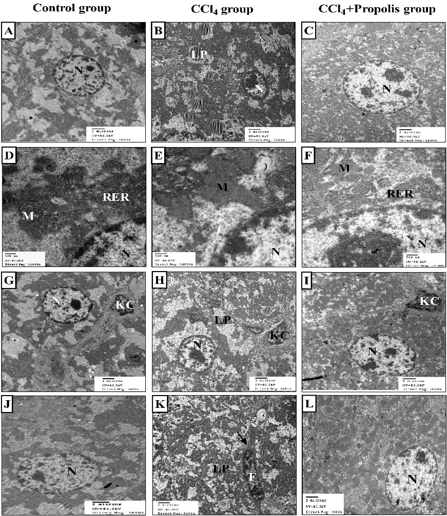
Fig. 5. Oral supplementation with propolis mitigated CCl4-mediated distortion of the hepatic architecture. Electron micrographs of liver sections from the three animal groups. Control group showing hepatocytes with a normal architecture of the nucleus (N) (A), CCl4-treated group showing hepatocytes with heterochromatic nucleus (B), and the presence of a large number of lipid droplets (LP). CCl4+propolis group showing hepatocytes with euchromatic nuclei (N) (3600X) (C). Control group showing part of a nucleus (N), rough endoplasmic reticulum (RER), and mitochondria (M) (D), CCl4-treated group showing part of a nucleus (N), devastated mitochondria (M) and fragmented rough endoplasmic reticulum (RER) (E), CCl4+propolis group showing part of the nucleus (N), with mitochondria (M), and rough endoplasmic reticulum (RER) localization near the nuclear envelope or scattered into the cytoplasm (14000X) (F). Control group showing hepatocytes with a normal nucleus (N), Kupffer cell (KC) (G), CCl4-treated group showing a nucleus (N) with increased heterochromatin patches, lipid droplets (LP), and Kupffer cell (KC) (H), CCl4+propolis group showing a nucleus (N) with normal chromatin content, and a Kupffer cell (KC) (3600X) (I). Control group showing a normal nucleus (N) (J), CCl4-treated group showing an elongated nucleus of fibroblast cell (F), some lipid droplets (LP), and collagen fibers (arrow) (K). CCl4+propolis group showing a normal-looking nucleus (N) of an hepatocyte (3600X) (L).